- 1 - Who is it for
- 2 - How to take
- 3 - Ingredients
- 4 - Frequently Asked Questions
Cartilage natural food supplement
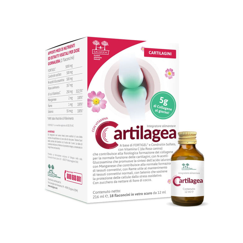
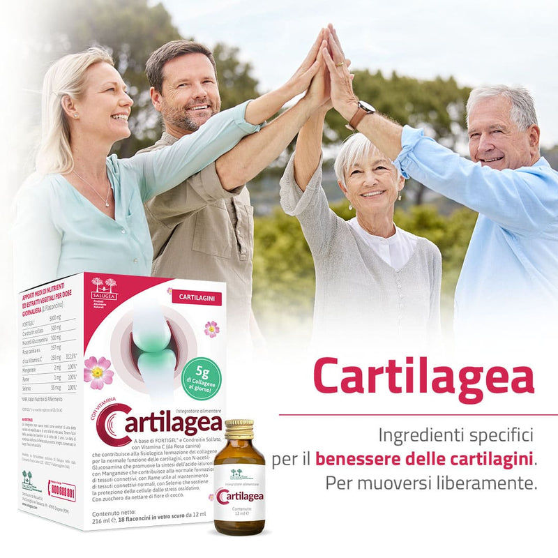
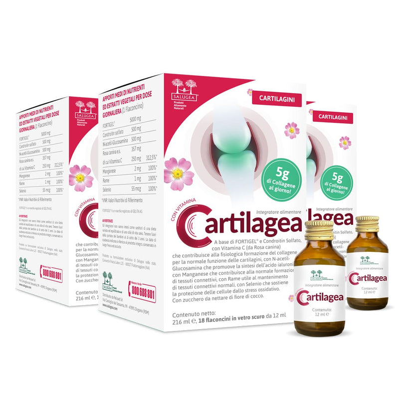
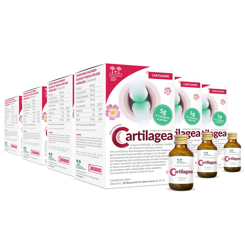
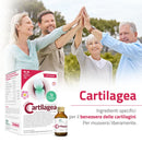
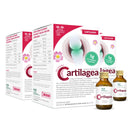
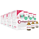
Cartilagea is Salugea 100% natural liquid supplement with specific natural ingredients that sustain the physiological functions of the joint cartilage and of the connective tissue of tendons and ligaments.
You will finally be able to move your body freely again, without pain nor effort, whatever your age!
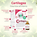
The efficacy of Cartilagea lies in its formulation! In fact, it provides the maximum allowable daily intake of Glucosamine and Chondroitin, which are essential to support the physiological functions of the cartilage and the joint connective tissue.
In addition, Cartilagea formulation also contains hydrolyzed collagen (FORTIGEL®, a patented extract that has been clinically proven to help the regeneration of cartilage cells*), Rosehip (Vit. C), as well as organic Manganese, Copper and Selenium.
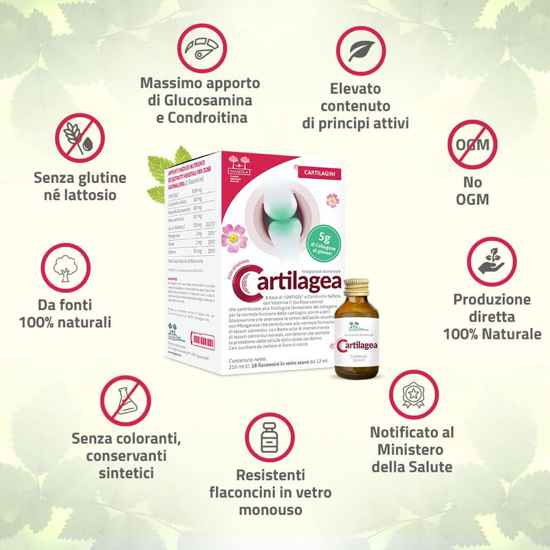
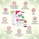
Cartilagea is packed in convenient, durable single-dose vials in pharmaceutical brown glass.
Free from dyes, preservatives and sweeteners.
A 100% natural, next-generation product with active ingredients exclusively obtained from 100% natural sources, therefore not synthetic (i.e. artificially made in labs). Besides, Cartilagea active ingredients work in perfect synergy for greater efficacy and longer-lasting effects.
It is a product obtained exclusively from natural sources that ensures high quality standards and a much greater assimilation.
Research confirms that natural molecules obtained from natural sources - which is the case with vegetable extracts - are far more easily assimilated by the human body than the synthetic ones (i.e. created in labs).
*See the Experimental Phytotherapy chapter below for the complete bibliography.
Who is Cartilagea for?
Cartilagea is recommended for those who:
- experience joint discomfort with movements,
- feel their joints stiff,
- stress their cartilage and joints intensely when doing sports,
- want to preserve the joint cartilage as they get older,
- prefer a natural, high-quality remedy for the cartilage,
- wish to support tendons and ligaments functions,
- tend to frequently have joint injuries.
What are the benefits you obtain thanks to Cartilagea active ingredients?
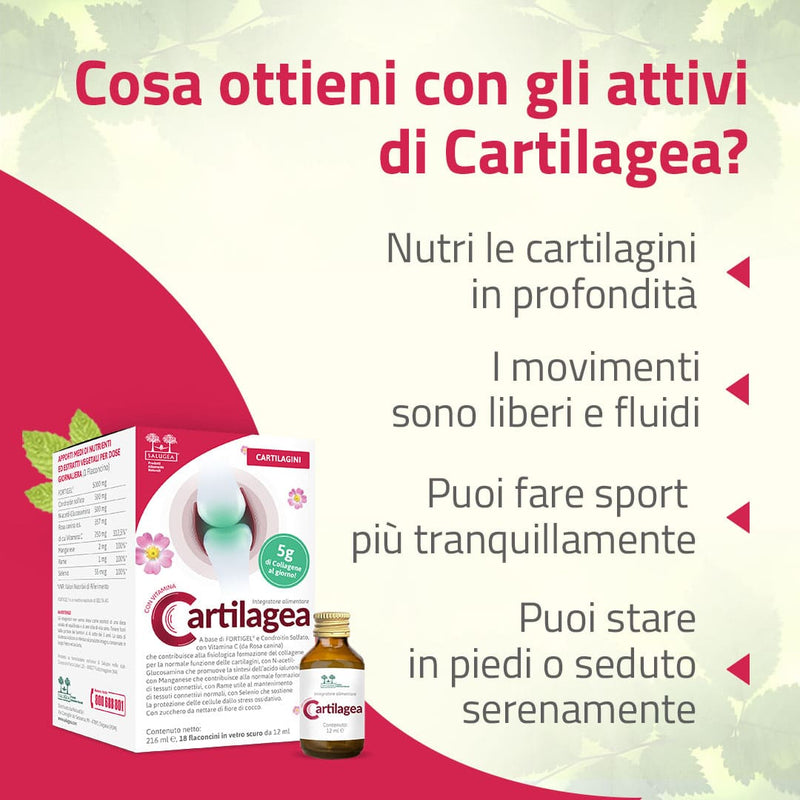
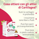
- Cartilage tissues are deeply nourished.
- You can move easily and freely again.
- You will be able to sit or stand with no more discomfort.
- You can enjoy your favorite sports with no more worries.
- Your joint cartilage, tendons and ligaments are sustained thanks to a complete and natural food supplement.
- The recovery of your tendons and ligaments after injury is supported.
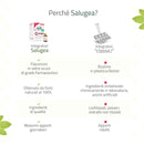
Why is Cartilagea different from any other cartilage supplement?
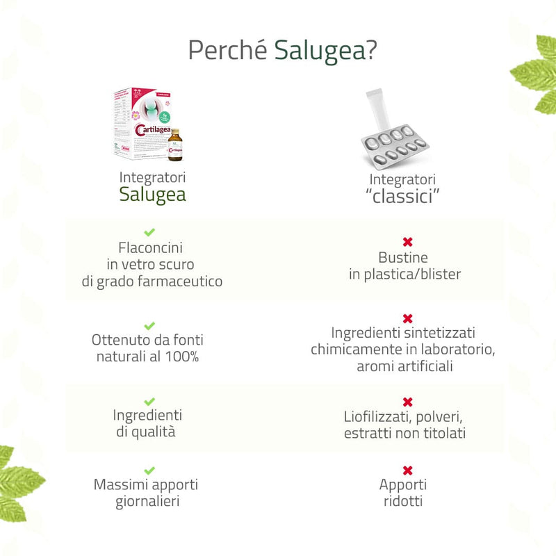
- It is packed in convenient and durable brown glass single-dose vials. Glass is the only material that can guarantee the quality and safety of the product over time, and it is good to the environment!
- It is 100% natural.
- This all-in-one supplement provides the maximum allowable daily intake of Glucosamine and Chondroitin.
- It also provides Hydrolyzed Collagen (FORTIGEL®), Rosehip (Vit. C), Manganese, Copper and Selenium in their organic form.
- Cartilagea active ingredients are all from 100% natural, non synthetic (i.e. made in labs) sources.
- Cartilagea formulation is innovative.
- It is gluten-free.
How Cartilagea works to sustain the joints cartilage
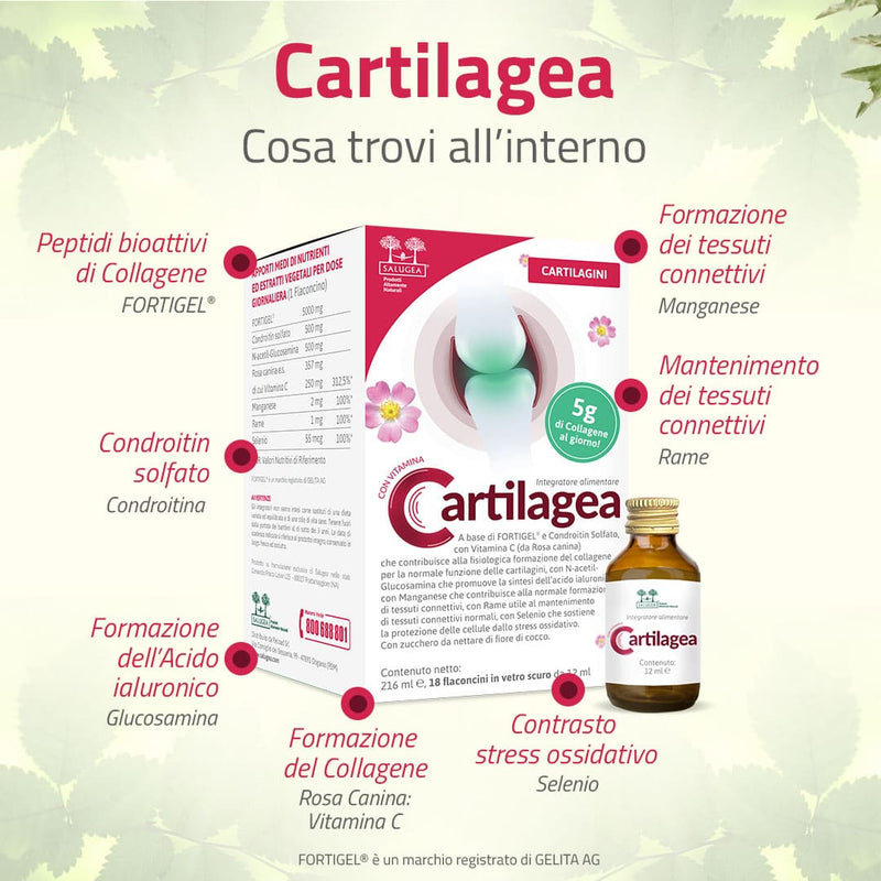
FORTIGEL® - bioactive collagen peptides - 5 g/day of collagen.
FORTIGEL® is an innovative ingredient that supports joint cartilage, as well as tendons and ligaments. Its bioactive peptides are easily absorbed by the intestines and their action and efficacy have been confirmed by numerous clinical studies*.
FORTIGEL® hydrolysed collagen peptides activate the growth of new cartilage tissue by stimulating the cells to produce cartilage matrix components. This helps recover joints flexibility and mobility*.
Chondroitin - 500 mg maximum allowable dose/day.
Chondroitin is essential to sustain the cartilage and joints physiological functions. In fact, it ensures the cartilage high elasticity and resistance to compression. Several studies* confirm the efficacy and benefits of oral administration of Chondroitin, especially on the hip and knee joints. Cartilagea contains the maximum allowable daily intake of natural Chondroitin.
Glucosamine - 500 mg maximum allowable daily intake, for the synthesis of hyaluronic acid.
Glucosamine helps keep cartilage intact and sustains tendons and ligaments. Scientific studies* demonstrate that it also plays an important role in the synthesis of new hyaluronic acid, a cartilage functional component of great importance. Cartilagea also contains the maximum allowable daily intake of natural Glucosamine.
Rosehip, for the supply of Vitamin C.
Vitamin C plays a key role in the synthesis of collagen, which is essential to keep the joint cartilage, tendons and ligaments in good condition. Rosehip is naturally rich in vitamin C. Cartilagea contains therefore a Vitamin C from an entirely natural source, unlike most of the similar products available on the market.
Manganese and Copper provide support to the connective tissues.
Their role is fundamental to sustain the joint cartilage function and structure. Particularly, Manganese helps the formation of the connective tissues whilst Copper stimulates the proliferation and differentiation of articular chondrocytes, the "load-bearing" cells of the cartilage*.
Selenium protects the cells from oxidative stress.
Selenium is an essential trace element which is beneficial to the whole body. It has been included in the formulation of Cartilagea because it can neutralise the effects of oxidative stress, which commonly affects the cartilage and articular tissue leading to its deterioration, shrinkage and loss of function.
For maximum benefits we recommend that you take 1 vial a day. Dilute in little water and preferably take away from meals. Shake well before use.
Duration of treatment with Cartilagea, the joint cartilage supplement.
You will soon see the results and feel the benefits. We advise you to carry on with the treatment for at least three months, so as to obtain the best results and keep them over time.
At the end of the treatment, please contact us! We care for you and would be most pleased to know how you got on with the treatment and how you feel. Our Team of experts (Biologists and Naturopathic Doctors) will advise you on the next steps to take in order to maintain the results you have achieved.
DISCLAIMER. As we are all different (and unique!), the dosage can be set according to your specific needs and requirements. Just give us a call or write to us for any kind of information or advice you may require. Our experts shall remain at your fullest disposal ;)
Taken alone, Cartilagea can be extremely effective. Nonetheless, for a stronger and comprehensive action, you may want to combine it with:
- Sanaos Nuova Formula, for a synergistic action supporting the bones and improving the joint mobility.
- Glutatione Forte, for an even greater antioxidative action, Glutathione being considered as one of the most powerful antioxidants in the world.
- Omega 3 Krill Oil, Superior Quality Omega 3 with no fishy aftertaste for a strong antioxidative action.
Warning
Supplements cannot be considered as a replacement for healthy diet and lifestyle. Keep out of reach of children under the age of three. Do not exceed the recommended daily intake. The expiry date refers to the product intact, stored in a cool and dry place, away from heat and direct sunlight.
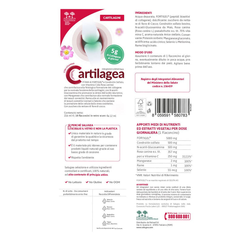
Purified water, FORTIGEL® (bioactive collagen peptides), sweetener: sugar from coconut flower nectar; bovine chondroitin sulfate, N-acetyl-Glucosamine from Maize, Rosehip (Rosa canina L.) pseudofruit d.e. tit. 70% Vitamin C, natural Anise flavor, Preservative: Potassium sorbate; Manganese gluconate, Selenium L-Methionine, Copper bisglycinate.
Nutritional Values
|
AVERAGE INTAKE OF NUTRIENTS AND VEGETABLE EXTRACTS PER DAILY DOSE (1 Vial) |
||
|
FORTIGEL® |
5'000 mg |
|
|
Chondroitin sulfate |
500 mg |
|
|
N-acetyl-Glucosamine |
500 mg |
|
|
Rosehip d.e. |
357 mg |
|
|
of which Vitamin C |
250 mg |
312,5% NRV |
|
Manganese |
2 mg |
100% NRV |
|
Copper |
1 mg |
100% NRV |
|
Selenium |
55 mcg |
100% NRV |
|
NRV: Nutrient Reference Values |
||
FORTIGEL® is a GELITA AG registered trademark.
Scientific Insights
Cartilage is a strong and flexible connective tissue that protects bones and joints by absorbing shocks and reducing friction during movement.
Cartilage contains metabolically active cells called chondrocytes and various kinds of structural proteins that vary in quantity according to each type of cartilage.
According to the composition of their tissue, cartilages can be of three types.
- Hyaline Cartilage is the most common type of cartilage that is found throughout the body. It lines the joints (articular cartilage), ribs, nose, trachea (windpipe), bronchi and larynx.
- Elastic cartilage is the most flexible cartilage in the body. Its locations in the body are, for instance, external ears (auricles) and epiglottis.
- Fibrocartilage is the strongest and least flexible of the three and is found in the meniscus of the knee, in disks between the spine vertebrae (intervertebral discs) and in pubic symphysis.
Cartilage is therefore a fundamental structure and has a peculiarity that makes it unique in the body: it is not vascularized (i.e. it has no blood vessels).
This characteristic entails that it is difficult for the cartilage to get nutrients and, at the same time, to get rid of toxins that tend to build up.
Another characteristic of the cartilage is that 65 to 80% of its weight is water.
Usually, cartilage problems appear after the age of 40 and are mainly due to wear and tear.
Articular cartilage injuries are often associated with other joint conditions. A study[1] on 200 cases showed that only 6.5% presented a single lesion, whilst 61.5% had multiple lesions.
Other similar structures at the joint level, which are also subjected to extreme stress, include tendons and ligament - fibrous connective elements composed of collagen and elastin.
Regenerative processes in tendons and ligaments are also very slow because these tissues are poorly vascularized and have a relatively low oxygen consumption. As a result, they are easily prone to injuries and micro-injuries caused by excessive effort or repeated strain (e.g., the Achilles tendon in runners, or tennis elbow in tennis players).
Natural remedies can do a great deal to support cartilage, tendons, and ligaments, and help people maintain a good quality of life by preserving their joints full functionality.
Let's see now what are the natural ingredients that have been included in Cartilagea based on scientific evidence.
Experimental Phytotherapy
FORTIGEL®
Clark KL et Al. 24-Week study on the use of collagen hydrolysate as a dietary supplement in athletes with activity-related joint pain. Curr Med Res Opin. 2008 May;24(5):1485-96. doi: 10.1185/030079908x291967. Epub 2008 Apr 15. PMID: 18416885.
This clinical study conducted at Penn State University (USA), involved 147 athletes experiencing activity-related joint pain. Athletes (aged 20.1 years on average) were divided into two groups. Over a period of 24 weeks, one group was administered FORTIGEL® (a dietary supplement), while the control group was administered a placebo. The severity of symptoms was assessed by both the treating physician and the athletes by means of an analog judgment scale. At the end of the 24-week treatment, athletes on FORTIGEL® showed an improvement on the symptomatology scale.
The study confirms that FORTIGEL® collagen hydrolysate administered to athletes can reduce discomfort (such as pain) thus improving their athletic performance.
McAlindon TE, Nuite M, Krishnan N, Ruthazer R, Price LL, Burstein D, Griffith J, Flechsenhar K. Change in knee osteoarthritis cartilage detected by delayed gadolinium enhanced magnetic resonance imaging following treatment with collagen hydrolysate: a pilot randomized controlled trial. Osteoarthritis Cartilage. 2011 Apr;19(4):399-405. doi: 10.1016/j.joca.2011.01.001. Epub 2011 Jan 18. PMID: 21251991.
The clinical study investigated FORTIGEL® long-term effects on the composition of hyaline cartilage, in individuals with early-stage knee osteoarthritis. Monitoring was carried out by MRI (magnetic resonance imaging) analysis of the cartilage. At the end of the study, subjects treated with FORTIGEL® - unlike the placebo group - showed a statistically significant increase in the density of proteoglycans in the medial and lateral tibial regions, which resulted in improved knee functions.
Chondroitin
Martel-Pelletier J, Kwan Tat S, Pelletier JP. Effects of chondroitin sulfate in the pathophysiology of the osteoarthritic joint: a narrative review. Osteoarthritis Cartilage. 2010 Jun;18 Suppl 1:S7-11. doi: 10.1016/j.joca.2010.01.015. Epub 2010 Apr 27. PMID: 20399897.
This review summarizes some data relating to the mechanisms of action of chondroitin sulfate in the pathophysiology of osteoarthritic joint tissues. Researchers suggest that chondroitin mechanism of action stimulates the chondrocytes synthesis of proteoglycans and decreases their catabolic activity by inhibiting the synthesis of proteolytic enzymes and other factors that contribute to the cartilage matrix damage and cause the chondrocytes death. Chondroitin sulfate has also been shown to exert a remarkable anti-inflammatory activity.
Wildi LM, Raynauld JP, Martel-Pelletier J, Beaulieu A, Bessette L, Morin F, Abram F, Dorais M, Pelletier JP. Chondroitin sulphate reduces both cartilage volume loss and bone marrow lesions in knee osteoarthritis patients starting as early as 6 months after initiation of therapy: a randomized, double-blind, placebo-controlled pilot study using MRI. Ann Rheum Dis. 2011 Jun;70(6):982-9. doi: 10.1136/ard.2010.140848. Epub 2011 Mar 1. PMID: 21367761; PMCID: PMC3086081.
The objective of the study was to determine the effect of chondroitin sulphate treatment on cartilage volume loss in patients with knee osteoarthritis. The six-month double-blind study involved 69 patients with clinical signs of synovitis. Cartilage volume was assessed by MRI (Magnetic Resonance Imaging). At the end of the research, the chondroitin sulphate group showed significantly less cartilage volume loss than the placebo group. These findings suggest that chondroitin sulphate has a protective effect on the joint structure of the knee affected by osteoarthritis which can be therefore successfully treated.
Glucosamine
Shikhman AR, Brinson DC, Valbracht J, Lotz MK. Differential metabolic effects of glucosamine and N-acetylglucosamine in human articular chondrocytes. Osteoarthritis Cartilage. 2009 Aug;17(8):1022-8. doi: 10.1016/j.joca.2009.03.004. Epub 2009 Mar 24. PMID: 19332174; PMCID: PMC2785807.
This study analyzes the metabolic effects of Glucosamine on articular chondrocytes in the knee cartilage. The results show that chondrocytes absorb Glucosamine which is used for the production of new hyaluronic acid. The increased synthesis of hyaluronic acid accelerated by Acetyl-Glucosamine is associated with the upregulation of the enzyme hyaluronan synthase-2, which confirms the functional action of this element.
Gallagher B, Tjoumakaris FP, Harwood MI, Good RP, Ciccotti MG, Freedman KB. Chondroprotection and the prevention of osteoarthritis progression of the knee: a systematic review of treatment agents. Am J Sports Med. 2015 Mar;43(3):734-44. doi: 10.1177/0363546514533777. Epub 2014 May 27. PMID: 24866892.
This work shows how the combined use of glucosamine and chondroitin sulfate is an effective non-surgical way to preserve the articular cartilage of the knee and delay the progression of osteoarthritis.
Rosehip (Vitamin C)
Sharma G, Saxena RK, Mishra P. Regeneration of static-load-degenerated articular cartilage extracellular matrix by vitamin C supplementation. Cell Tissue Res. 2008 Oct;334(1):111-20. doi: 10.1007/s00441-008-0666-9. Epub 2008 Aug 5. Erratum in: Cell Tissue Res. 2009 May;336(2):347. PMID: 18679720.
This in-vitro study investigated the effect of a physiological dose of vitamin C on chondrocytes. The results show that in-vitro vitamin C supplementation has the potential to reduce the degeneration of chondrocytes, thereby improving the cellular health of articular cartilage.
Chang Z, Huo L, Li P, Wu Y, Zhang P. Ascorbic acid provides protection for human chondrocytes against oxidative stress. Mol Med Rep. 2015 Nov;12(5):7086-92. doi: 10.3892/mmr.2015.4231. Epub 2015 Aug 20. PMID: 26300283.
The study aimed to assess the effects of Vitamin C on human chondrocytes. Evidence showed that ascorbic acid increases the production of collagen and proteins and inhibits the differentiation of chondrocytes under conditions of oxidative stress, thus promoting the preservation of cartilage tissue.
Manganese, Copper and Selenium
Valero G, Alley MR, Badcoe LM, Manktelow BW, Merrall M, Lawes GS. Chondrodystrophy in calves associated with manganese deficiency. N Z Vet J. 1990 Dec;38(4):161-7. doi: 10.1080/00480169.1990.35645. PMID: 16031605.
This study was carried out on calves affected by a severe cartilage malformation (congenital chondrodystrophy). It was found that the feed given to pregnant cows was very low in Manganese, which caused a histological deficiency in the calves’ cartilage.
Pasqualicchio M, Gasperini R, Velo GP, Davies ME. Effects of copper and zinc on proteoglycan metabolism in articular cartilage. Mediators Inflamm. 1996;5(2):95-9. doi: 10.1155/S0962935196000154. PMID: 18475704; PMCID: PMC2365780.
Copper plays a strategic role in the physiological preservation of cartilage. This in-vitro study investigated the effect of Copper and Zinc on the chondroblasts capability of producing proteoglycans, in a porcine articular cartilage. It was observed that Zinc exerts no stimulating action on chondroblasts, whilst Copper does. This finding was supported by the histological demonstration of copper-dependent reversal of the proteoglycan depletion in cartilage.
Kang D, Lee J, Wu C, Guo X, Lee BJ, Chun JS, Kim JH. The role of selenium metabolism and selenoproteins in cartilage homeostasis and arthropathies. Exp Mol Med. 2020 Aug;52(8):1198-1208. doi: 10.1038/s12276-020-0408-y. Epub 2020 Aug 13. PMID: 32788658; PMCID: PMC7423502.
The study investigates the link between Selenium deficiency and selenoproteins dysregulation that are associated with impaired redox homeostasis in cartilage leading to the onset and progression of osteoarthritis, and similar pathological conditions.
Cartilage and tendons
Nutraceuticals containing multiple specific nutrients can be helpful in managing discomfort affecting both cartilage and tendons[2] [3], thanks to the anti-inflammatory effects of some natural ingredients and their support of the body’s natural connective tissue repair processes.
Therefore, supplements composed of, for example, collagen peptides, chondroitin, vitamin C, and similar components may have strong potential as an additional approach alongside standard treatment methods (such as physical exercise or rehabilitation)[4] [5].
DISCLAIMER. The content is not intended to be a substitute for professional medical advice, diagnosis, or treatment. Always seek the advice of your physician or other qualified health provider with any questions you may have regarding your medical condition.
[1] Articular cartilage lesions of the knee. Ronald W. Zamber, M.D. Carol C. Teitz, M.D. David A. McGuire, M.D. John D. Frost, M.D.
[2] Hijlkema A, Roozenboom C, Mensink M, Zwerver J. The impact of nutrition on tendon health and tendinopathy: a systematic review. J Int Soc Sports Nutr. 2022 Aug 3;19(1):474-504. doi: 10.1080/15502783.2022.2104130. PMID: 35937777; PMCID: PMC9354648.
[3] Burton I, McCormack A. Nutritional Supplements in the Clinical Management of Tendinopathy: A Scoping Review. J Sport Rehabil. 2023 May 5;32(5):493-504. doi: 10.1123/jsr.2022-0244. PMID: 37146985.
[4] Choudhary A, Sahu S, Vasudeva A, Sheikh NA, Venkataraman S, Handa G, Wadhwa S, Singh U, Gamanagati S, Yadav SL. Comparing Effectiveness of Combination of Collagen Peptide Type-1, Low Molecular Weight Chondroitin Sulphate, Sodium Hyaluronate, and Vitamin-C Versus Oral Diclofenac Sodium in Achilles Tendinopathy: A Prospective Randomized Control Trial. Cureus. 2021 Nov 19;13(11):e19737. doi: 10.7759/cureus.19737. PMID: 34812335; PMCID: PMC8603329.
[5] Dos Santos DR, Xavier DP, de Ataíde LAP, Bentes LGB, Lemos RS, Giubilei DB, de Barros RSM. The Effects of Hydrolyzed Collagen and Collagen Peptide in the Treatment of Superficial Chondral Lesions: An Experimental Study. Rev Bras Ortop (Sao Paulo). 2022 Oct 20;58(1):72-78. doi: 10.1055/s-0042-1756332. PMID: 36969779; PMCID: PMC10038713.
I nostri testi hanno scopo divulgativo, non vanno intesi come indicazione di diagnosi e cura di stati patologici e non vogliono sostituirsi in alcun modo al parere del Medico.
Cod. Min. Sal.: 156439
I nostri testi hanno scopo divulgativo, non vanno intesi come indicazione di diagnosi e cura di stati patologici e non vogliono sostituirsi in alcun modo al parere del Medico.
Cartilagea
Cartilage food supplement
Domande Frequenti sull'integratore
Altre Domande?
Se non hai trovato la risposta alla tua domanda, puoi contattarci:

